Anatomical Characteristics of Trigger Points:-
- The trigger point is a hard, tender "lump" in the body of a muscle. It may be as small as a pinpoint or as big as your thumb, and is exquisitely tender to pressure from finger or thumb or tennis ball.
- The trigger point is characterized by intense contractile activity in the absence of nerve excitation. (Like a muscle with a cramp, but in a small circumscribed area).
- The trigger point is not an inflamed area of muscle; very few microscopic investigations of trigger points reveal the presence of inflammatory cells(1). (But refer discussion, bottom of page). However, severe damage at the Sub-cellular and molecular levels has been noted, and will be now be discussed.
Trigger Point Diagram 1: A Trigger Point in a Muscle
Note the bunching up of the muscle fibers in the trigger point knot, and the stretching and elongation of the muscle fibers to either side of the trigger point. 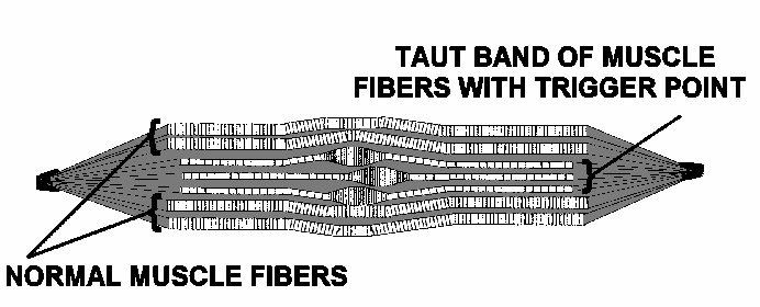
Trigger Point Micro-anatomy: Severe damage at the Sub-cellular and Molecular Levels.
|
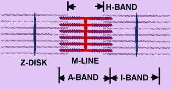
Trigger Point Diagram 2: The Sarcomere - Molecular unit of Muscle Contraction. Intense contractile activity in the area of a trigger point results in tearing of the actin molecules in the region of the Z-disk (adapted from Michael W. King: Muscle Biochemistry - ref. 11).
 Trigger Point Diagram 3: The Myosin and Actin Molecules - "Ropes" that walk along each other. Note the blue "legs" on the Myosin molecule. During contraction, these "legs" walk along the Actin molecule. Each Myosin unit can have in excess of 200 "legs". 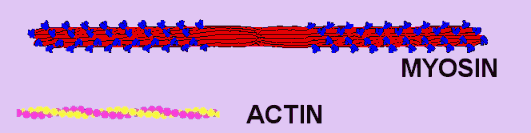 |
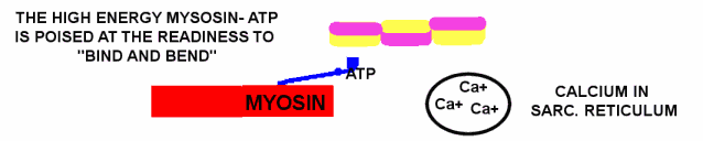
Muscles contract when, in response to a motor nerve signal, Calcium is released from storage in the sarcoplasmic reticulum. The next two diagrams and captions discuss this.
Trigger Point Diagram 4: Resting Muscle: In the resting muscle, ATP is bound to Myosin in a high energy configuration. The Myosin cannot however do anything until Calcium is released from the sarcoplsamic reticulum. The Calcium switch is turned off.
Trigger Point Diagram 5: Muscle in Rigor Mortis - the actin-myosin unit is locked together and cannot release: The Calcium switch has turned on, and has enabled the high energy Myosin-ATP to "bind and bend" to the Actin with the release of low energy ADP and Phosphate. However that is as far as it goes: there is no more energy currency (ATP) in the muscle cell to drive the Myosin cycle of "release and straighten, bind and bend", and the Myosin remains stuck fast to the actin molecule.

Muscle with Trigger Point Dysfunction:
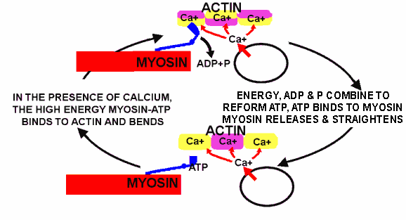
Trigger Point Diagram 6: Active Muscle - In this active muscle, both Calcium and ATP are present, and the Myosin cycle of "Release and straighten, bind and bend" is in full flight. The myosin is "walking up the actin". Normal muscle has sufficient ATP in the cell and sufficient integrity in its sarcoplasmic reticulum to quickly re-absorb the Calcium and turn off the contraction. However, a trigger point zone in a muscle cannot re-absorb the Calcium and turn the contraction off: The contraction continues until tension pulls hard enough against the Myosin "leg" to stop it at the bending phase of its cycle. Small wonder that there is intense activity with build up of lactic acid and molecular damage.
Clinical Application: Why Can't the Calcium Switch in the Trigger Point Zone turn off?
This is no idle question. If we knew why the Calcium switch fails to turn off, we could potentially develop better therapeutic strategies. Here are some suggestions, alongside therapeutic implications:-
- Mechanical stress to the Sarcoplasmic reticulum: Bunching of the sarcoplasmic reticulum may break its structural integrity -
Stretching, or even resting a muscle at a slightly longer length may enhance sufficient Calcium switch integrity to let it turn off. Likewise avoidance of unnecessary prolonged pressure, for example the "fat wallet" in the back pocket (a common cause of trigger point mediated buttock pain).
- Depletion of Cellular energy and build up of Lactic Acid: Bunching of muscle tissue at the trigger point zone severely disables blood flow, and this, alongside the energy intensive Actin-Myosin activity reduces the cellular energy (ATP) available to the Calcium switch. It also increases local levels of lactic acid, which can be detrimental to cellular function -
Massage, or any gently continuous movement is likely to encourage better blood supply and therefore more available energy at the cellular level, also dietary Minerals, B group vitamins and malic acid may enhance cellular energy metabolism. Also, a reduction in carbohydrates (who's metabolism requires large quantities of B group vitamins) may benefit.
- Poor membrane integrity of the sarcoplasmic reticulum due to "toxic" (hydrogenated and heavily oxidised polyunsaturate) fats: Leads to a poorly functional and easily damaged Calcium switch -
Replace these types of fats with healthy ones in the diet, and increase the dietary anti-oxidants.
- Statins: Disrupt the production of Co-enzyme Q-10, and also the normal and natural membrane component "Cholesterol" -
Muscle pain and/or weakness is one of the many side effects of Statins. If you have these symptoms, consider discontinuing the drug class "Statins", because they destroy Co-enzyme Q-10, which is a very important lipid based anti-oxidant, and because the manufacture of the (highly necessary and completely normal) cholesterol component of sarcoplasmic reticulum membranes.
More information, trigger point precipitating factors, see "Trigger Point Diagram Cascade for Diagnosis and Management", reference 12.
Final Note Re: Inflammation at the Trigger Point Zone
An Injection of "aspirin like" anti-inflammatory (Diclofenac) into a trigger point relieves pain more strongly than an injection of local anesthetic (1). So there is good evidence for prostaglandin release at the trigger point zone. Evidence for other inflammatory or nerve pain substances (substance P, Leukotrienes, Histamine, Serotonin) at the trigger point site is more controversial.
These substances, together with signs of microscopic inflammation are however to be found at zones of neurogenic inflammation. For more information, refer: Biology of Trigger Points page 9: Neurogenic Inflammation
'건강하고 행복하게 > 建康 運動' 카테고리의 다른 글
| 인체구조와 운동방법 (0) | 2010.03.04 |
|---|---|
| 運動과 呼吸 WEIGHT TRAINING AND RESPIRATION (0) | 2009.12.13 |
| Adenosine triphosphate (0) | 2009.11.20 |
| Biological Energy - ADP & ATP (0) | 2009.11.20 |
| The Amazing Energy Cycle: ATP-ADP (0) | 2009.11.20 |