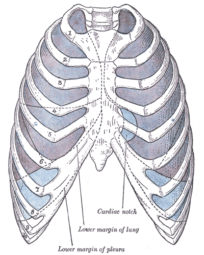
In human anatomy, the pleural cavity is the body cavity that surrounds the lungs. The lungs are surrounded by the pleurae, a serous membrane which folds back upon itself to form a two-layered, membrane structure. The thin space between the two pleural layers is known as the pleural space; it normally contains a small amount of pleural fluid. The outer pleura (parietal pleura) is attached to the chest wall. The inner pleura (visceral pleura) covers the lungs and adjoining structures, i.e. blood vessels, bronchi and nerves.
The parietal pleura is highly sensitive to pain; the visceral pleura is not due to its lack of sensory innervation.[1]
Functions
The pleural cavity, with its associated pleurae, aids optimal functioning of the lungs during respiration. The pleurae are coated with lubricating pleural fluid which allows the pleurae to slide effortlessly against each other during ventilation. Surface tension of the pleural fluid also leads to close apposition of the lung surfaces with the chest wall. This physical relationship allows for optimal inflation of the alveoli during respiration. Movements of the chest wall, particularly during heavy breathing, are coupled to movements of the lungs since the closely opposed chest wall transmits pressures to the visceral pleural surface and, hence, to the lung itself
Pleural fluid
Pleural fluid is a serous fluid produced by the pleurae. In normal pleurae, most fluid is produced by the parietal circulation (intercostal arteries) via bulk flow and reabsorbed by the lymphatic system. Thus, pleural fluid is produced and reabsorbed continuously. In a normal 70 kg human, a few milliliters of pleural fluid is always present within the intrapleural space.[2] Larger quantities of fluid can accumulate in the pleural space only when the rate of production exceeds the rate of reabsorption. Normally, the rate of reabsortion increases as a physiological response to accumulating fluid, with the reabsorption rate increasing up to 40x before significant amounts of fluid accumulate within the pleural space. Thus, a profound increase in the production of plural fluid, or some blocking of the reabsorbing lymphatic system, is required for fluid to accumulate in the pleural space.
When accumulation of pleural fluid is noted, cytopathologic evaluation of the fluid, inclusive of total cell counts and protein determination, is often recommended as a diagnostic tool for determining the causes of accumulation. The presence of heart failure, infection or malignancy within the pleural cavity are the most common causes that can be identified using this approach.[3]

'연구하는 인생 > 西醫學 Medicine' 카테고리의 다른 글
| inflammation 의 종류 (0) | 2009.02.19 |
|---|---|
| Healing (0) | 2009.02.16 |
| ☆ 당뇨병 과 운동 ☆ (0) | 2009.01.03 |
| DIABTETS - BRITANNICA (0) | 2009.01.01 |
| Alzheimer's disease (0) | 2008.12.19 |